Dental Pulp: Morphology, Histology, Innervation, Vascular Supply, Neurotransmitters, Development and Functions
Dental Pulp
Morphology of Pulp
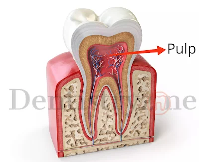
The shape of the dental pulp is the same as the crown of the tooth which is present. The pulp is present in the crown and the root portion has coronal pulp and radicular pulp.
The extension of the pulp chamber near the cusp tips is called pulp horns. The radicular portion of the pulp is seen in the inner surface of the root known as the root canal. A Root may have one or more root canals. If lateral branches are present in the canals are called accessory canals.
Histology of Pulp
Under a microscope, the pulp consists of four zones. They are
i) Odontoblastic Zone
ii) Cell-free zone of Weil
iii) Cell-rich zone
iv) Pulp core
These zones are not seen as prominent in the radicular portion of the pulp.
Odontoblastic Zone
Odontoblasts are arranged in this zone surrounding the pulp adjacent to the unmineralized dentin called prevention. The number of odontoblasts is more abundant in coronal pulp than the radicular pulp. These odontoblast cells have the extension called the odontoblastic process. The process extends into the dentinal tubules up to dentinoenameljunction (DEJ). The fluid is present between the odontoblast process and the dentinal tubules. The fluid transmits the cold and hot stimuli to the pulp through the odontoblastic process which causes sensitivity.
Cell-free zone of Weil
It is located between the odontoblastic zone and cell-rich zone. There are no cells seen in the cell-free zone of Weil. Some fibres run through this zone. Some research studies suggest that this zone permits odontoblasts inwards to the pulp.
Cell rich zone
This layer of the contains rich in contains fibroblasts and blood vessels. The fibroblasts maintain the pulp matrix. It degrades and synthesizes collagen fibres. It secretes the extracellular components such as ground substances and proteoglycans. It also secretes Type-I, Type-III, Type-V and Type-VI collagens.
Apart from the fibroblast, the cell-rich zone contains undifferentiated mesenchymal cells. It is a totipotent stem cell which may be differentiated into odontoblast, fibroblast and macrophages when the demand arises.
The defence cells are normal habitants of the dental pulp. Macrophages, Lymphocytes, plasma cells, neutrophils, eosinophils, and monocytes are also seen in the pulp. They respond to dental caries, chemical irritants, and mechanical irritants.
Pulp Core
The pulp core mainly comprises the ground substances which occupy the extracellular spaces. Collagen, Proteoglycans, and Glycoproteins are the major components of the pulp core.
Vascular Supply of Pulp
The blood pressure and blood flow of the dental pulp are higher than in other tissues in the body. Two vessels of 150 Micrometres enter the pulp through the apical foramen accompanied by sensory and sympathetic nerve bundles. The vessels get bigger in the coronal portion of the pulp. Some lateral branches arise from the coronal vessels of the pulp which would not carry any nerve bundles. Lymphatic vessels are present in the pulp.
Innervation of the pulp
Pulp is innervated by sensory and autonomic nerve endings.
Sensory Nerves
These nerves are involved in the sensation of pain and transduction. Sensory nerves are derived from the maxillary and mandibular branch of trigeminal nerves which reaches the pulp through the apical foramen. They are branched to form the parietal layer of nerves or plexus of Raschkow near the cell-rich zone. The free endings of the sensory nerves reach the odontoblastic layer and penetrate the predentin and form the specific receptors for pain.
Autonomic sympathetic nerve
The sympathetic nerve fibres originate from the cervical sympathetic ganglion and combine with the trigeminal nerve at its ganglion. The sympathetic vasoconstriction is stimulated by the pain and stress which modulates the excitability of sensory nerves.
Neurotransmitters of the pulp
Substance P, 5-hydroxytryptamine, vasoactive intestinal peptide, somatostatin, prostaglandin, acetylcholine and norepinephrine are the neurotransmitters within the pulp. These neurotransmitters affect the vascular tone and excitability of the nerve endings.
The result of all external stimuli such as
heat, touch, pressure and chemicals are a pain.
Development of the pulp
The development of the pulp is started in the 8th week of intrauterine life. The dental papilla is referred to as the dental pulp once the first layer of calcified matrix is deposited during the bell stage of tooth development which is called predentin. Undifferentiated mesenchymal cells in the dental papilla differentiate into fibroblasts. The neural innervations are also initiated. Plexus of Raschkow is not formed till root completion. The lifespan of deciduous pulp is 8 years and 3 months.
Functions of the pulp
Inductive
The pulp helps in the induction of the dental papilla cells to form the enamel
Formative
The pulp has odontoblasts which cause the formation of the dentin. It also secretes the organic matrix for dentin formation.
Nutritive
The pulp has highly vascular tissue. It provides nutrients to the odontoblasts.
Protective
Cells such as monocytes, macrophages, lymphocytes, neutrophils, plasma cells and mast cells try to act as the defence cells to protect the pulp.
Reparative
The pulp overcomes the injury to dental caries, mechanical irritants, heat, cold, and chemical irritants by the formation of the sclerotic dentin.


COMMENTS