What is an Oral Mucous Membrane? The lining of the oral cavity is called as oral mucous membrane. It consists of epithelium and ...
What is an Oral Mucous Membrane?
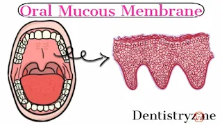
The lining of the oral cavity is called as oral mucous membrane. It consists of epithelium and connective tissue. The connective tissue of the Oral mucosa is known as lamina propria.
Classification of Oral Mucous Membrane
Based on functional criteria,
- Masticatory Mucosa
- Lining Mucosa
- Specialized mucosa
Masticatory Mucosa
It is found on the gingiva and hard palate. It helps to withstand the forces during mastication. It contributes about 25% of the total oral mucosa. It is lined by keratinized squamous epithelium. It has both ortho-keratinized wells as para-keratinized.
Lining mucosa
It is also known as reflecting mucosa. It is found in the lip, cheek, alveolar mucosa, vestibular fornix, the floor of the mouth and soft palate. It contributes about 60℅ of the oral mucosa. It is lined by non-keratinized squamous epithelium
Specialized mucosa
It is present in the dorsum of the tongue and lateral surface of the tongue. It contributes 15℅ of the oral mucosa. It is lined by a keratinized epithelium.
Based on the type of epithelial covering,
- Keratinized mucosa
- Non-keratinized mucosa
Keratinized mucosa
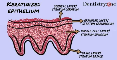
This mucosa contains keratins. Histologically, the epithelium of keratinized mucosa has four layers. They are,
- Stratum basale
- Stratum spinosum
- Stratum granulosum
- Stratum corneum
An example of keratinized mucosa is the hard palate, gingiva, vermilion border of the lip and some papillae of the tongue.
Non-keratinized mucosa
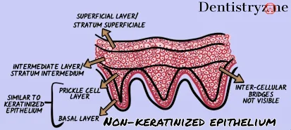
This mucosa cannot have keratins. Histologically, the epithelium of non keratinized mucosa has three layers. They are,
- Stratum basale
- Stratum intermedium
- Stratum superficial
Examples of non-keratinized mucosa are lining mucosa and some parts of the gingiva.
Functions of the oral mucous membrane
Defense
If the continuity of the oral epithelium happens, the bacteria and other microorganisms in the oral cavity attack the damaged site then the infection occurs. So structural integrity of oral epithelium acts as an effective barrier for invasion of the microorganisms.
Protection
It helps in the protection of the tissues by mechanical forces which results due to mastication.
Lubrication
The oral mucous membrane has several salivary glands which help to prevent the dryness of the oral cavity and maintaining moisture. The presence of moisture is essential for better speech, mastication, Swallowing and perception of taste functions.
Sensory
The sensation of taste is unique and it is present in the anterior 2/3rd of the tongue. And it is sensitive to pain, hot and cold temperatures.
Histological features of keratinized mucosa (keratinized stratified squamous epithelium)
As I said earlier, it is made up of four layers.
Stratum basale
This is the basal layer of the epithelium, beneath this layer connective tissue called "lamina propria" is present. This basal layer connects the epithelium to the underlying connective tissue with the help of a protein called hemidesmosomes. Hemidesmosomes are more in number and larger.
Stratum spinosum
This the layer just above the basal layer. Cells in this layer are polygonal in shape and show a prickle appearance. This layer is also known as the prickle cell layer. Desmosomes are helping to bind the adjacent cells in this layer. In this layer, cell size is small. Cytokeratin present here is 1, 6, 10, 16. Cytokeratins bundled to form the Tonofilaments. Odland bodies are present in the layer. It is also known as lamellar granules or keratinosomes.
Stratum granulosum
The cells in this layer are flat and bigger than the spinous layer. Keratohyalin granules are seen in this layer. This layer is also known as "Granular Layer".
Stratum corneum
This layer contains tightly packed tonofilaments(contains cytokeratin). Cells in this layer do not have a nucleus and cytoplasmic organelles. This layer is also known as "Corneal Layer".
Histological features of Non-keratinized mucosa (keratinized stratified squamous epithelium)
The histological section of this layer shows only three layers.
Stratum basale
This layer is similar to the keratinized epithelium. But the inter-cellular bridges i.e, Hemidesmosomes are not visible as compared to the keratinized epithelium.
Stratum Intermediate
The cells in this layer are bigger than the granular layer of keratinized epithelium but have dispersed tonofilaments and not aggregated as tonofibrils. Lamellate granules are circular with the amorphous core. These cells also secrete lipid material into intercellular spaces between the intermediate and superficial layer but it is found not effective barrier compared to the keratinized epithelium. Intermediate cells may sometimes have keratohyalin granules.
Stratum superficial
Cells in this layer have a flat nucleus. Unlike the corneal layer of keratinized epithelium, the cells in this layer have a nucleus and other cells organelles. Tonofilaments are dispersed in the cell. Cells withstand the masticatory forces.


COMMENTS